


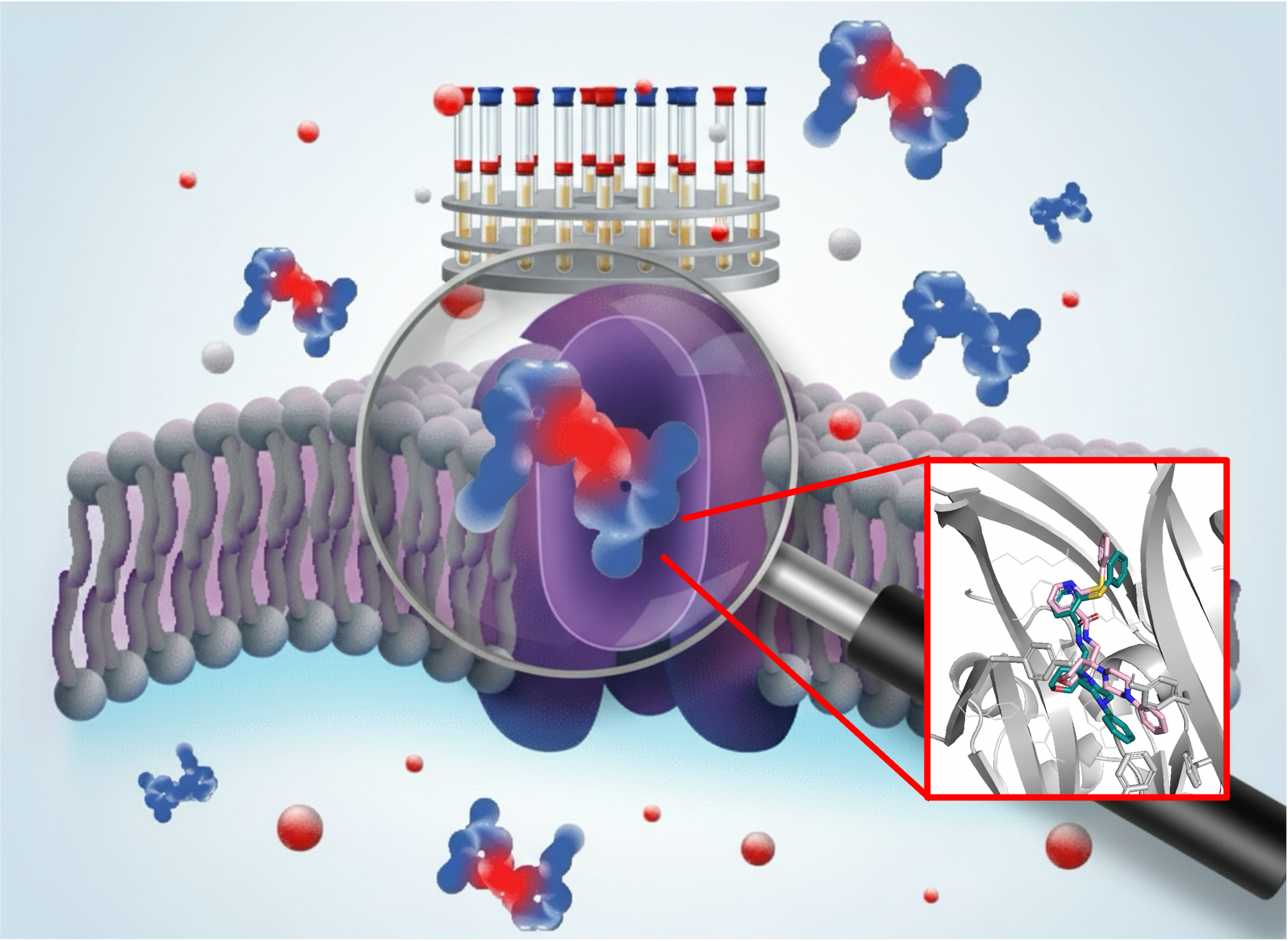
An international team with the participation of the Spanish National Research Council (CSIC) has developed a new technique that will make it possible to accelerate the design of drugs targeting ion channels, a type of cell membrane protein involved in numerous diseases, ranging from psychiatric disorders to various types of cancer. The research— a collaboration between the Spanish National Research Council (CSIC), the University of East Anglia, and the Quadram Institute (both in the United Kingdom)—has been published in the Journal of the American Chemical Society.
Ion channels are cell membrane proteins that regulate the flow of ions into the cell and are fundamental to processes as diverse as nerve transmission, muscle contraction, and the immune response. Their dysfunction is associated with numerous pathologies, making them highly attractive therapeutic targets.
“Until now, studying how drugs interact with these proteins required isolating them, a technically complex process that can alter their behavior. Our technique, based on nuclear magnetic resonance, allows these interactions to be studied in living cells, providing biologically more relevant information,” explains Jesús Angulo, from the Institute of Chemical Research (a joint center of CSIC and the University of Seville).
The new technique is faster—based on experiments lasting less than an hour—more cost-effective, and significantly simpler, as it eliminates the need for complex prior protein purification processes or sample manipulation.
The researchers believe that their method could become a standard tool for structure–activity studies aimed at understanding how the chemical structure of a molecule relates to its pharmacological effect.
“Our technique could significantly accelerate the development of drugs targeting ion channels and other membrane proteins, opening up new research possibilities in multiple areas, from neurological or cardiovascular diseases to metabolic and oncological disorders,” notes Leanne Stokes of the University of East Anglia, United Kingdom.
A new tool for pharmacological studies
The new technique has been tested on P2X7 receptors, ion channels that are therapeutic targets for depression, certain autism spectrum disorders, and some types of cancer. “We have shown that we can identify, directly in living cells, which parts of the drug interact with the protein, allowing us to optimize these interactions—information that is essential for developing more effective and specific medicines,” says Serena Monaco, a researcher at the Quadram Institute in Norwich, also in the United Kingdom.
In addition, thanks to software developed at the IIQ-CSIC-US, the authors combined these experimental data with three-dimensional models of drug–receptor binding generated through bioinformatics, enabling them to validate which computer-proposed models actually matched what was observed in the laboratory.
“The interaction between a drug and a protein can be compared to a key and a lock. The membrane protein is the lock and our key is the drug. But not only do we need to find the right key; we also need to determine how to insert it so that it opens more efficiently,” illustrates Angulo. “Bioinformatic models are key to designing new drugs. Being able to validate three-dimensional computational models directly in living cells represents a new paradigm in the development of drugs targeting these proteins,” concludes the CSIC researcher.